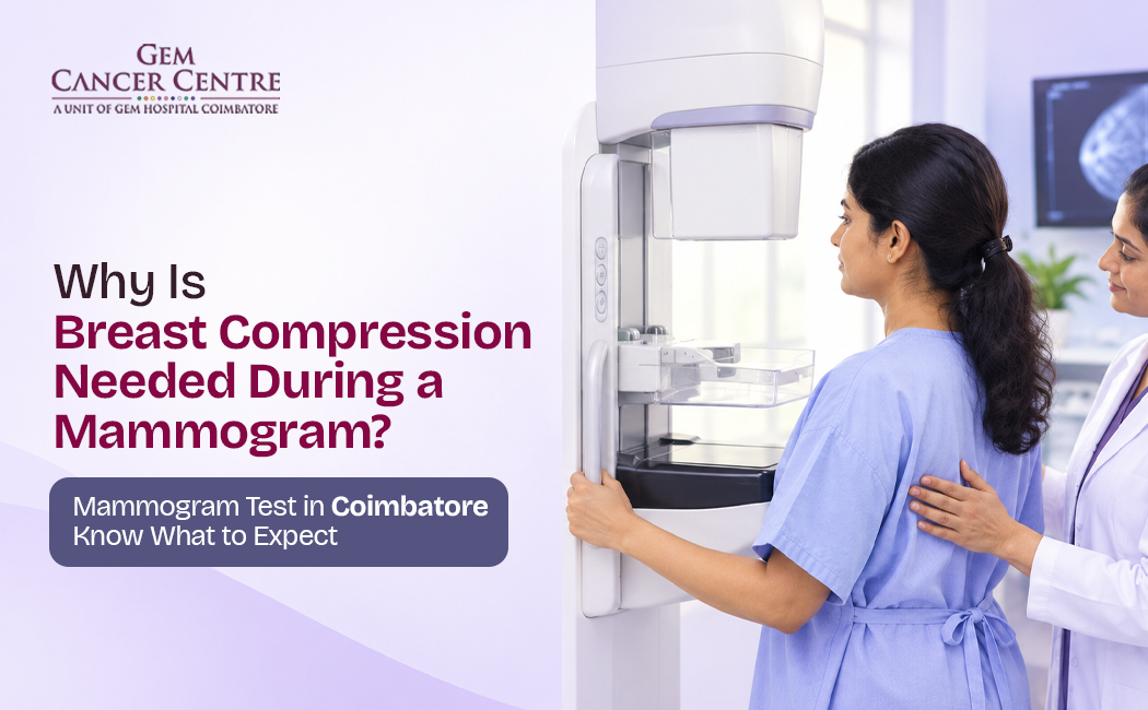
If you are preparing for a Mammogram test in Coimbatore, you may have questions about one part of the procedure that many women find uncomfortable: breast compression.
During a mammogram, the breast is gently but firmly compressed between two plates for a few seconds while X-ray images are taken. Although compression can feel uncomfortable, it plays an important role in obtaining clear and useful images of breast tissue.
Breast compression is not intended to cause harm. It helps the radiologist examine breast tissue more clearly and can improve the ability to identify small abnormalities that may not be easy to see otherwise.
Understanding why compression is necessary can make the mammogram experience less stressful.
What Is Breast Compression During a Mammogram?
Breast compression gently spreads breast tissue for clearer X-ray images.
During a mammogram, the breast is positioned between two compression plates. The plates gradually apply pressure for a short period while the image is captured.
The amount of compression varies depending on the breast and the image being taken.
Compression is a standard part of mammography because it helps produce images with greater clarity.
Why Is Breast Compression Needed During a Mammogram?
Compression improves image quality and can reduce radiation exposure.
There are several important reasons for compressing the breast during mammography.
1. It Spreads Out Breast Tissue
Breast tissue can overlap when viewed on an X-ray image.
Compression spreads the tissue into a thinner, more uniform layer. This can make it easier for the radiologist to distinguish normal structures from areas that may need further evaluation.
2. It Helps Detect Small Abnormalities
When breast tissue is spread out, abnormalities such as small masses or calcifications may be easier to identify.
This is particularly important in screening, where the purpose is to look for changes that may not yet be causing symptoms.
3. It Reduces Blurring
Even small movements during imaging can affect the clarity of an X-ray.
Compression helps keep the breast stable while the image is captured, reducing motion-related blurring.
4. It Can Reduce Radiation Dose
A thinner layer of compressed breast tissue requires less X-ray exposure to create a useful image.
Therefore, appropriate compression can help produce a diagnostic-quality image while limiting radiation exposure.
Does Breast Compression Hurt?
Compression can feel uncomfortable, but it lasts only seconds.
Women experience mammogram compression differently.
Some may describe it as pressure or tightness, while others may find it temporarily uncomfortable.
The compression is applied only for the short period needed to capture each image.
If the pressure becomes excessively painful, tell the mammography technologist. Communication during the examination is important.
Does More Compression Mean a Better Mammogram?
Appropriate compression matters more than maximum pressure.
The goal is not simply to apply as much pressure as possible.
The breast needs to be compressed sufficiently to spread the tissue, reduce movement and create a good-quality image.
The mammography technologist determines appropriate positioning and compression according to the examination.
How Does Compression Improve Mammogram Images?
Compression separates overlapping breast tissue for better visibility.
Imagine trying to examine several transparent sheets stacked on top of each other. It can be difficult to see what is on each individual sheet.
Breast tissue can create a similar challenge on an X-ray image.
Compression spreads the tissue, helping reduce overlap and making structures easier to evaluate.
This is one reason why proper positioning and compression are important parts of a mammogram.
Is Breast Compression Safe?
Yes, mammogram compression is generally considered safe.
Compression is a routine part of mammography and is used to obtain appropriate images.
Some women may experience temporary tenderness or discomfort after the examination, but significant lasting effects from routine mammographic compression are not expected.
If you have recently had breast surgery, an injury or significant breast tenderness, inform the mammography team before the examination.
Can Compression Cause Breast Cancer?
No, routine mammogram compression does not cause breast cancer.
This is a common concern among women who have never undergone mammography.
The purpose of compression is to improve the quality of the X-ray image. It does not cause cancer.
Mammography uses a low dose of X-ray radiation and the benefits of appropriate screening should be discussed with a healthcare professional based on age, risk factors and individual circumstances.
What Happens During a Mammogram Test?
A typical mammogram involves several simple steps.
Step 1: Registration and Preparation
You may be asked to remove clothing and jewellery from the upper body and change into a gown.
Avoid applying deodorant, talcum powder or certain cosmetic products around the breast and underarm area before the examination if instructed by the imaging centre, as some products can create marks on mammogram images.
Step 2: Breast Positioning
The technologist positions one breast at a time on the mammography unit.
Proper positioning is important because it helps include as much relevant breast tissue as possible in the image.
Step 3: Compression
The breast is compressed between the plates.
The pressure is maintained for a few seconds while the image is captured.
Step 4: Additional Views
More than one image of each breast is usually taken.
Additional images may sometimes be required depending on breast anatomy or the purpose of the examination.
Step 5: Image Review
The images are reviewed by an appropriately trained radiologist.
If an area requires further evaluation, additional imaging may be recommended.
Can You Prepare for a Mammogram?
Simple preparation can make the examination easier.
Before your appointment:
Wear comfortable clothing.
Avoid creams or powders around the breasts if instructed.
Tell the technologist if you are experiencing breast tenderness.
Inform the imaging team if you may be pregnant.
Bring previous mammogram reports or images if available.
Follow any specific instructions provided by the imaging centre.
There is generally no need for fasting for a routine mammogram unless another test is being performed at the same appointment.
Is a Mammogram Uncomfortable for Everyone?
No, discomfort varies from person to person.
Breast sensitivity can differ depending on factors such as hormonal changes, individual pain sensitivity and breast tenderness.
Some women find mammography easier when scheduled at a time when their breasts are less tender.
If you are particularly anxious or have experienced painful mammograms before, discuss your concerns with the mammography team.
What Is the Difference Between a Screening and Diagnostic Mammogram?
Screening checks for cancer signs; diagnostic imaging investigates concerns.
A screening mammogram is generally performed in people without breast symptoms as part of breast cancer screening.
A diagnostic mammogram may be recommended when there is a symptom, a clinical finding or an abnormality that needs closer assessment.
Diagnostic imaging may involve additional mammographic views or other breast imaging depending on the situation.
Why Is Mammogram Screening Important?
Breast cancer can sometimes develop without causing noticeable symptoms in its early stages.
Mammography can help detect certain breast changes before they become apparent through physical symptoms.
However, mammography is not perfect. Dense breast tissue and other factors can affect how clearly abnormalities appear.
Your doctor may recommend additional imaging depending on your age, breast density, symptoms and individual risk.
When Should You Consider a Mammogram?
Screening recommendations depend on age and individual risk.
There is no single screening schedule suitable for every woman.
Your healthcare professional may consider:
Age
Family history
Previous breast conditions
Genetic risk
Personal medical history
Previous imaging findings
Women with a strong family history or other high-risk factors may require a different screening strategy.
If you notice a breast lump, nipple discharge, skin changes, nipple inversion or another unusual change, do not wait for a routine screening appointment. Seek medical evaluation.
What If the Mammogram Shows an Abnormality?
An abnormal mammogram does not automatically mean cancer.
Many findings on mammography are benign.
If something unusual appears, the doctor may recommend additional tests such as:
Additional mammographic views
Breast ultrasound
Breast MRI in selected situations
Biopsy when clinically necessary
The purpose of additional testing is to understand the finding more accurately.
Where Can You Get a Mammogram Test in Coimbatore?
When choosing a centre for a Mammogram test in Coimbatore, consider factors such as:
Availability of appropriate mammography equipment
Qualified radiology professionals
Proper breast positioning and imaging technique
Clear reporting
Access to additional breast imaging when required
Appropriate referral for further evaluation
A good mammography experience should focus not only on obtaining images but also on accurate interpretation and appropriate follow-up.
Frequently Asked Questions
1. Why is breast compression required during a mammogram?
Breast compression spreads the tissue, reduces overlap and movement, improves image quality and can help reduce the amount of radiation needed.
2. Is mammogram compression painful?
Compression can be uncomfortable for some women, but the pressure is usually applied only for a few seconds during each image.
3. Can breast compression damage breast tissue?
Routine mammogram compression is designed to safely hold and spread the breast tissue while images are taken. Significant tissue damage is not expected.
4. Does compression increase the risk of breast cancer?
No. Compression itself does not cause breast cancer. Mammography uses a low dose of X-ray radiation to create breast images.
5. Why can't a mammogram be done without compression?
Without adequate compression, overlapping tissue and movement can make the images less clear and may reduce the ability to identify small abnormalities.
6. Does compression reduce radiation exposure?
Yes. By making the breast thinner and reducing tissue overlap, appropriate compression can help produce a useful image with a lower X-ray exposure than would otherwise be required.
7. How long does breast compression last?
The compression for each image generally lasts only a few seconds, although the overall examination takes longer because multiple images may be required.
8. Should I tell the technician if compression hurts?
Yes. Tell the mammography technologist if you experience significant pain or discomfort. Good communication can help make the examination more comfortable.
9. Can I have a mammogram if I have a breast lump?
Yes, but a breast lump should be medically evaluated. Depending on your age and symptoms, your doctor may recommend diagnostic mammography, ultrasound or other tests.
10. Is a mammogram enough to detect every breast cancer?
No test detects every breast cancer. Mammography is an important breast-imaging tool, but additional imaging or biopsy may be required depending on symptoms, breast density and imaging findings.
Conclusion
Breast compression is one of the most important parts of a mammogram. Although the pressure may feel uncomfortable for a few seconds, it helps spread breast tissue, reduce movement, improve image quality and potentially lower the radiation needed to obtain useful images.
If you are planning a Mammogram test in Coimbatore, understanding the purpose of compression can make the procedure feel less unfamiliar.
Most importantly, do not let fear of compression prevent you from discussing breast screening with your healthcare professional. The appropriate timing and type of breast imaging depend on your age, symptoms and individual risk factors.
Call to Action
If you have been advised to undergo a mammogram or have concerns about breast health, speak with a qualified healthcare professional and choose an imaging centre where you can receive appropriate breast imaging and expert interpretation.
Early evaluation can provide clarity when breast changes are detected and help guide the next step in care.